
Seeing Beyond the Surface: How Modern Dentistry Has Changed
Imagine taking your car to a mechanic because of a strange noise coming from the engine. Now imagine the mechanic trying to diagnose the problem without ever opening the hood. Most of us would immediately question how accurate that diagnosis could possibly be.
The same principle applies to dentistry.
Many dental problems develop beneath the surface, hidden from the naked eye. A tooth may appear healthy on the outside while an infection quietly spreads within the bone. A wisdom tooth may seem harmless until its position threatens neighboring teeth. A patient considering dental implants may have excellent-looking gums, yet insufficient bone volume to support long-term success.
For decades, dentists relied primarily on conventional two-dimensional X-rays to investigate these conditions. While traditional radiographs remain extremely valuable, they provide a limited view of complex three-dimensional structures. Important details can sometimes overlap, remain hidden, or appear differently than they actually exist.
Today, advances in digital technology have transformed the way we diagnose and plan dental treatment. One of the most significant innovations in modern dentistry is Cone Beam Computed Tomography, more commonly known as a CBCT scan.
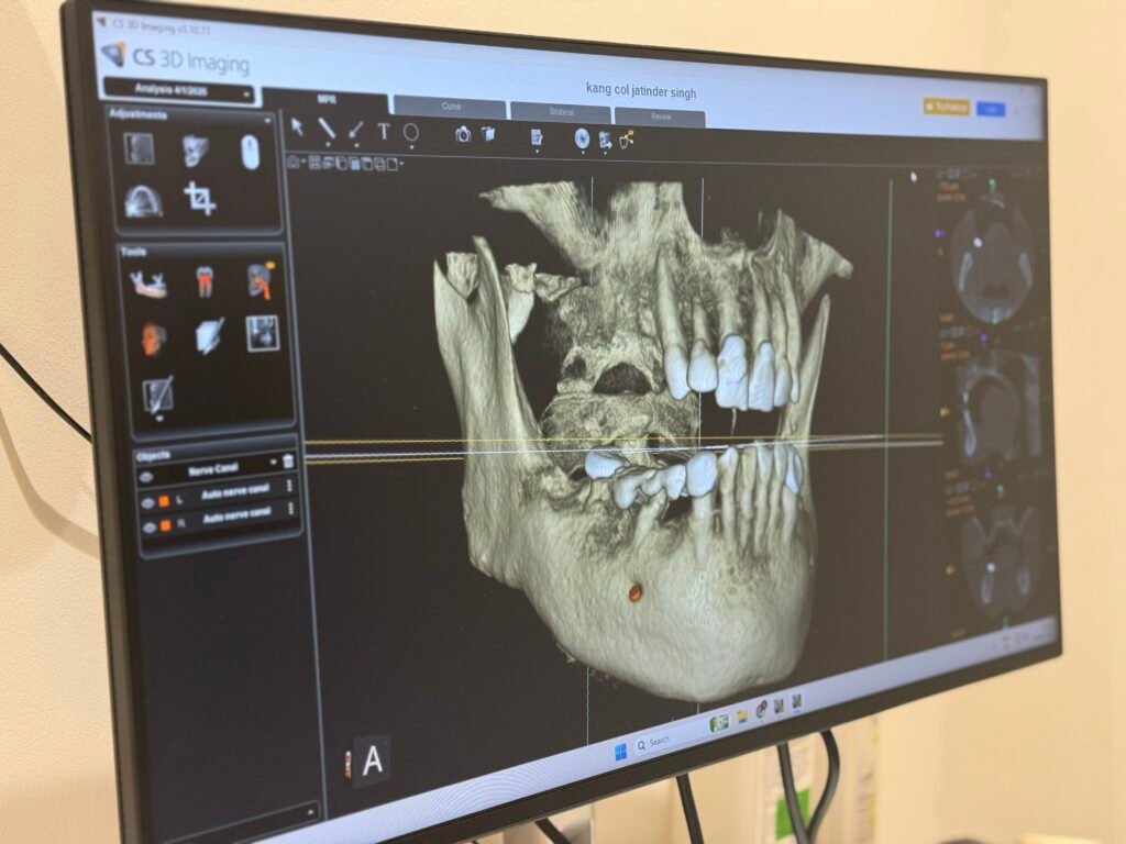
A CBCT scan allows dentists to view your teeth, jawbones, nerves, airways, sinuses, and surrounding structures in three dimensions, providing a level of detail that conventional dental X-rays simply cannot offer. It helps us move beyond educated assumptions and toward accurate, evidence-based diagnosis.
At Gelos Dentistry, we believe that the quality of treatment begins with the quality of diagnosis. The more precisely we understand a problem, the more confidently we can recommend solutions that are safe, predictable, and tailored to your individual needs.
What Exactly Is a CBCT Scan?
CBCT stands for Cone Beam Computed Tomography.
Although the name may sound intimidating, the concept is quite simple.
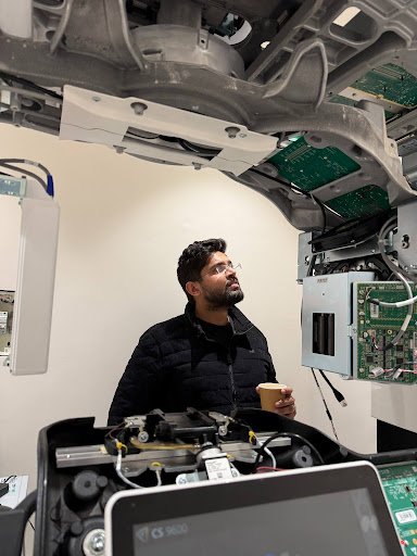
A CBCT scanner uses a specialised cone-shaped X-ray beam that rotates around your head while capturing hundreds of images from different angles. Advanced software then combines these images into a detailed three-dimensional representation of your teeth, jaws, facial structures, and surrounding anatomy.
Unlike a traditional dental X-ray, which produces a flat image, a CBCT scan allows your dentist to examine structures from multiple perspectives. We can rotate the image, view it layer by layer, zoom into specific areas, and evaluate anatomical details with exceptional clarity.
Think of it as the difference between looking at a photograph of a building and walking through a detailed 3D model of that same building. Both provide information, but one offers a much deeper understanding.
This enhanced visualisation helps dentists identify conditions that might otherwise remain hidden and allows treatment planning to be carried out with a much higher degree of confidence.
Why 2-Dimensional Images Sometimes Aren’t Enough
Traditional dental radiographs remain an essential part of modern dental care. They are fast, effective, and suitable for many routine situations.
However, the human face is a three-dimensional structure.
When a three-dimensional object is compressed into a flat image, certain details may overlap or become difficult to interpret. This limitation becomes particularly important when evaluating complex cases involving bone, nerves, impacted teeth, root canal anatomy, or implant placement.
For example, imagine trying to determine the exact position of an impacted wisdom tooth located near an important nerve. A standard X-ray may provide a general idea, but it cannot always reveal the precise spatial relationship between the two structures.
Similarly, when planning dental implants, understanding bone width, height, density, and anatomical limitations is critical. Even a small miscalculation can influence treatment outcomes.
CBCT technology eliminates much of this uncertainty by allowing dentists to visualise these structures in three dimensions before treatment begins.
How Does a CBCT Scan Work?
One of the most common concerns patients have is whether the procedure is complicated or uncomfortable.
The good news is that the entire process is remarkably simple.
During the scan, you will either sit or stand comfortably while the scanner rotates around your head. The machine never encloses you in a tunnel, making the experience much more comfortable than many patients expect.
The scan itself typically takes less than a minute.
There are no injections, no special preparations, and no recovery time afterwards. Once the scan is completed, the images are processed digitally and become available for detailed evaluation.
Patients are often surprised by how quick and straightforward the process feels compared to what they initially imagined.
What Can a CBCT Scan Reveal?
One of the greatest strengths of CBCT technology is the amount of information it provides.
The scan allows dentists to evaluate:
Teeth
Individual teeth can be examined in extraordinary detail. Hidden infections, fractures, root anatomy, impacted teeth, and developmental abnormalities become much easier to identify.
Jawbone Structure
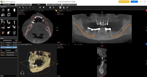
Bone volume, density, and quality can be accurately assessed before procedures such as dental implants or oral surgery.
Nerves
Important nerve pathways can be visualised clearly, helping reduce risks during surgical procedures and implant placement.
Sinuses
The relationship between teeth and the maxillary sinuses can be evaluated, which is particularly useful when planning implants in the upper jaw.
Impacted Teeth
Wisdom teeth and other impacted teeth can be assessed precisely to determine their position and relationship to surrounding structures.
Airway Analysis
In selected cases, CBCT imaging can also provide valuable information about airway dimensions and anatomical structures associated with breathing.
This comprehensive view helps clinicians understand not just isolated teeth but the entire oral and facial environment.
When Might Your Dentist Recommend a CBCT Scan?
Not every dental appointment requires a CBCT scan. In many situations, conventional dental X-rays provide all the information necessary for diagnosis and treatment planning.
However, certain cases demand a deeper understanding of the underlying anatomy. When greater precision is needed, a CBCT scan can provide valuable information that helps improve treatment outcomes and reduce uncertainty.
Your dentist may recommend a CBCT scan when planning dental implants, evaluating impacted wisdom teeth, diagnosing complex root canal problems, assessing jawbone conditions, investigating facial pain, planning orthodontic treatment, or examining areas that are difficult to visualise using traditional radiographs.
Rather than being a routine test, a CBCT scan serves as a powerful diagnostic tool when additional information can directly influence clinical decision-making.
CBCT Scans and Dental Implant Planning
Dental implants have transformed the way we replace missing teeth. However, successful implant treatment depends heavily on careful planning before the procedure begins.
When placing an implant, the dentist must evaluate several critical factors, including:
- Available bone height
- Bone width
- Bone density
- Position of nearby nerves
- Relationship to adjacent teeth
- Sinus anatomy in the upper jaw
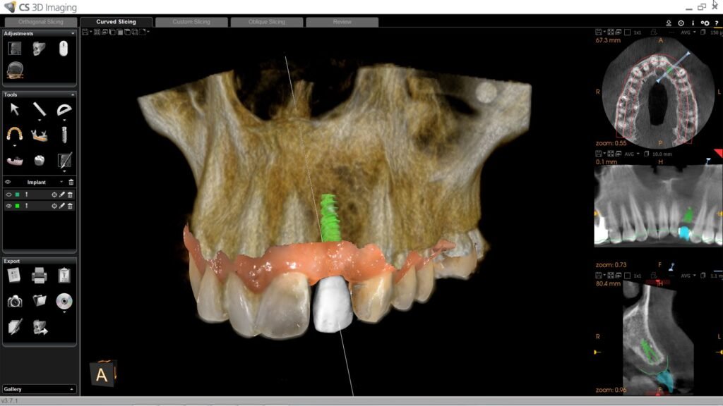
A CBCT scan allows all of these structures to be evaluated in three dimensions before implant placement.
Instead of estimating bone availability, the dentist can measure it precisely. This level of accuracy improves treatment predictability and helps create a surgical plan tailored specifically to the patient’s anatomy.
For patients considering dental implants, CBCT imaging often serves as one of the most important steps in the planning process.
How CBCT Improves Root Canal Treatments
Many patients assume that every tooth contains a single straight root canal. In reality, root canal anatomy can be remarkably complex.
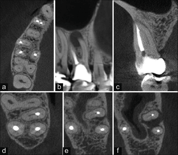
Some teeth contain multiple canals. Others have curved pathways, accessory branches, or unusual anatomical variations that may not be visible on conventional radiographs.
When a patient experiences persistent symptoms after previous treatment, a CBCT scan can often reveal hidden causes such as:
- Missed canals
- Undetected infections
- Root fractures
- Resorption defects
- Complex root anatomy
By providing a three-dimensional view of the tooth and surrounding bone, CBCT imaging helps clinicians identify problems more accurately and plan treatment with greater confidence.
For endodontists and dentists performing microscope-assisted root canal treatment, CBCT imaging has become an invaluable diagnostic resource.
Evaluating Wisdom Teeth with Greater Precision
Wisdom teeth frequently present unique challenges because they often develop in positions that are difficult to assess using conventional imaging alone.
Some wisdom teeth remain partially erupted. Others become trapped beneath the gums or bone. In certain cases, their roots develop close to important nerves that supply sensation to the lower lip and chin.
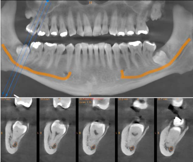
A CBCT scan allows the dentist or oral surgeon to evaluate:
- The exact position of the wisdom tooth
- Root development
- Bone coverage
- Relationship to neighboring teeth
- Proximity to vital nerves
This information helps clinicians plan safer extractions and discuss potential risks more accurately with patients before treatment begins.
The Role of CBCT in Orthodontic Treatment
Straightening teeth involves much more than simply moving them into alignment.
Successful orthodontic treatment requires a comprehensive understanding of jaw relationships, bone support, facial structures, airway dimensions, impacted teeth, and overall dental development.
In selected cases, CBCT imaging can assist in evaluating:
- Impacted canines
- Jaw discrepancies
- Skeletal relationships
- Airway anatomy
- Tooth positioning
- Root alignment
By providing a complete three-dimensional assessment, CBCT technology supports more informed treatment planning and helps orthodontists make decisions that consider both aesthetics and long-term function.
Beyond Teeth: Understanding the Bigger Picture
One of the most fascinating aspects of CBCT imaging is its ability to reveal structures beyond the teeth themselves.
The scan can provide valuable information about:
- Jaw joints (TMJ)
- Airway anatomy
- Sinus structures
- Facial bones
- Skeletal asymmetries
This broader perspective allows clinicians to view oral health within the context of overall facial anatomy rather than focusing solely on individual teeth.
In many situations, understanding these relationships can significantly influence diagnosis and treatment recommendations.
Is a CBCT Scan Safe?
Safety is understandably one of the most common questions patients ask when discussing any form of radiographic imaging.
Modern CBCT technology is specifically designed to capture detailed diagnostic information while using carefully controlled radiation exposure.
Dentists do not recommend CBCT scans unnecessarily. Instead, they are prescribed only when the additional information obtained is expected to improve diagnosis, treatment planning, or patient outcomes.
The guiding principle in modern dentistry is to use imaging responsibly and only when the benefits clearly outweigh the risks.
If a CBCT scan is recommended, your dentist should explain why it is being advised and how the information will contribute to your care.