
How Dental Microscopes Improve Root Canal Treatment Success
When Millimetres Make The Difference Between Saving and Losing a Tooth
One of the most common misconceptions about root canal treatment is that it is a straightforward procedure. Many patients imagine the inside of a tooth as a simple hollow space that can be cleaned and sealed with ease.
The reality is very different.
Inside every tooth lies a remarkably intricate network of canals, branches, curves, and microscopic anatomical variations. Some canals are so narrow that they can be difficult to identify even under bright clinical lighting. Others may split unexpectedly, curve sharply, or remain hidden from view entirely.
When these anatomical details go undetected, they can become a source of persistent infection, post-treatment discomfort, or treatment failure.
This is precisely where modern technology has transformed endodontics.
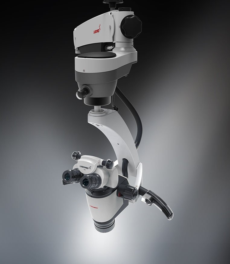
The introduction of dental operating microscopes has changed the way dentists diagnose, perform, and evaluate root canal treatment. By providing exceptional magnification and illumination, microscopes allow clinicians to visualize details that would otherwise remain invisible to the naked eye.
At Gelos Dentistry, we believe that precision forms the foundation of successful dentistry. By integrating advanced technology into clinical practice, we aim to provide patients with a higher standard of care and more predictable treatment outcomes.
Understanding the Challenge of Root Canal Treatment
A root canal procedure involves removing infected or damaged tissue from within the tooth, disinfecting the internal canal system, and sealing the space to prevent future bacterial contamination.
While the concept appears simple, the anatomy inside a tooth rarely is.
Many teeth contain multiple canals.
Some teeth contain additional accessory canals.
Others have extremely narrow passages that may not be visible during conventional treatment.
Research has consistently demonstrated that untreated canals remain one of the most common reasons for root canal failure. If bacteria remain hidden within an untreated area of the canal system, infection can persist even after treatment appears complete.
Successful endodontic treatment therefore depends not only on clinical skill but also on the ability to identify and treat every relevant part of the root canal system.
What Is a Dental Operating Microscope?
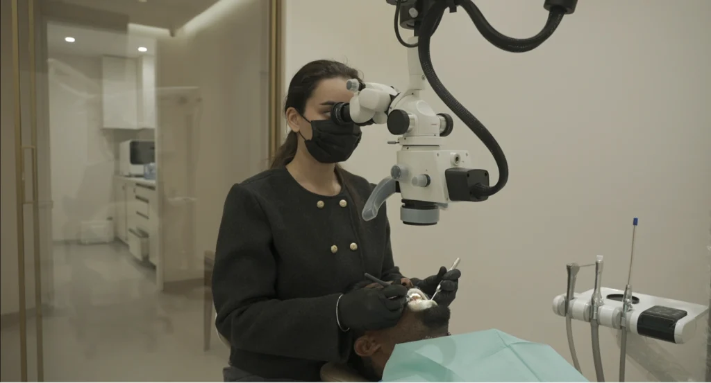
A dental operating microscope is a highly specialised optical instrument designed to magnify treatment areas while providing intense, focused illumination.
Unlike magnifying glasses or loupes, microscopes can provide significantly greater levels of magnification, allowing clinicians to visualise microscopic details within teeth and surrounding structures.
The microscope does not perform the treatment itself.
Rather, it allows the dentist to work with a level of visibility that was previously impossible.
Think of it this way:
If standard vision allows you to see a road, microscope-assisted dentistry allows you to see individual pebbles on that road.
The difference may seem subtle, but in dentistry, tiny details often determine long-term success.
Why Visibility Matters in Endodontics
Many dental procedures take place within confined spaces.
Root canals can be narrower than a fraction of a millimetre.
Cracks may be microscopic.
Hidden canals can remain concealed beneath layers of dentin.
Without adequate magnification, identifying these structures becomes significantly more challenging.
By improving visualisation, dental microscopes allow clinicians to:
- Locate additional canals
- Identify missed anatomy
- Detect cracks and fractures
- Remove obstructions
- Preserve healthy tooth structure
- Improve treatment precision
Greater visibility ultimately leads to better decision-making throughout treatment.
Finding Hidden Canals Before They Cause Problems
One of the most valuable advantages of microscope-assisted dentistry is the ability to locate hidden canals.
Many teeth contain anatomical variations that differ from textbook descriptions.
For example, upper molars frequently contain an additional canal known as the MB2 canal. This canal often remains difficult to locate during conventional treatment yet may harbour bacteria capable of sustaining infection.
Under magnification, clinicians can identify subtle anatomical clues that help reveal these hidden structures.
Finding and treating these canals significantly improves the likelihood of long-term success.
Detecting Cracks Before They Become Catastrophic
Not all tooth pain originates from infection.
Sometimes a patient experiences discomfort because of microscopic cracks that develop within the tooth structure.
These fractures may be impossible to identify with the naked eye, particularly during their early stages.
Dental microscopes allow clinicians to inspect teeth at much higher magnification, improving the detection of cracks, craze lines, and structural defects.
Early diagnosis often helps guide treatment decisions and may prevent more extensive complications in the future.
Improving Precision During Treatment
Precision matters in every area of healthcare.
In dentistry, precision often determines how much healthy tooth structure can be preserved.
The microscope allows clinicians to perform treatment in a more conservative and controlled manner.
Instead of removing excessive tooth structure to improve visibility, dentists can rely on magnification to work within smaller, more precise treatment areas.
This minimally invasive approach helps preserve the natural strength of the tooth while maintaining treatment effectiveness.
Managing Complex Cases with Greater Confidence
Some patients arrive after previous root canal treatment has failed.
Others present with unusual anatomy, calcified canals, broken instruments, or persistent infections.
These situations often require a higher level of diagnostic and clinical precision.
Microscope-assisted endodontics allows clinicians to evaluate complex cases more thoroughly and perform procedures that may otherwise prove significantly more difficult.
For patients seeking retreatment, this enhanced visibility can play an important role in identifying the cause of ongoing symptoms.
The Combination of CBCT and Dental Microscopes
Modern endodontics achieves its greatest potential when advanced technologies work together.
A CBCT scan allows dentists to visualise the tooth and surrounding anatomy in three dimensions before treatment begins.
The microscope then allows clinicians to apply that information during treatment with exceptional precision.
Together, these technologies create a powerful diagnostic and treatment workflow.
At Gelos Dentistry, we often combine advanced imaging and magnification to improve diagnosis, treatment planning, and clinical outcomes.
Does Microscope-Assisted Root Canal Treatment Feel Different for Patients?
One question patients frequently ask is whether microscope-assisted treatment feels different from conventional root canal therapy.
The answer is generally no.
Patients do not feel the microscope itself.
The primary difference occurs behind the scenes.
The microscope allows the clinician to work with greater accuracy and confidence while maintaining a comfortable patient experience.
Most patients simply appreciate knowing that advanced technology is being used to improve the quality of their treatment.
Common Situations Where a Dental Microscope Is Particularly Valuable
Microscope-assisted dentistry offers significant benefits in situations such as:
Complex Root Canal Anatomy
Retreatment of Failed Root Canal Cases
Detection of Hidden Canals
Identification of Cracks and Fractures
Removal of Broken Instruments
Management of Calcified Canals
Minimally Invasive Dentistry
Precision Restorative Procedures
In each of these scenarios, improved visibility contributes directly to clinical decision-making and treatment accuracy